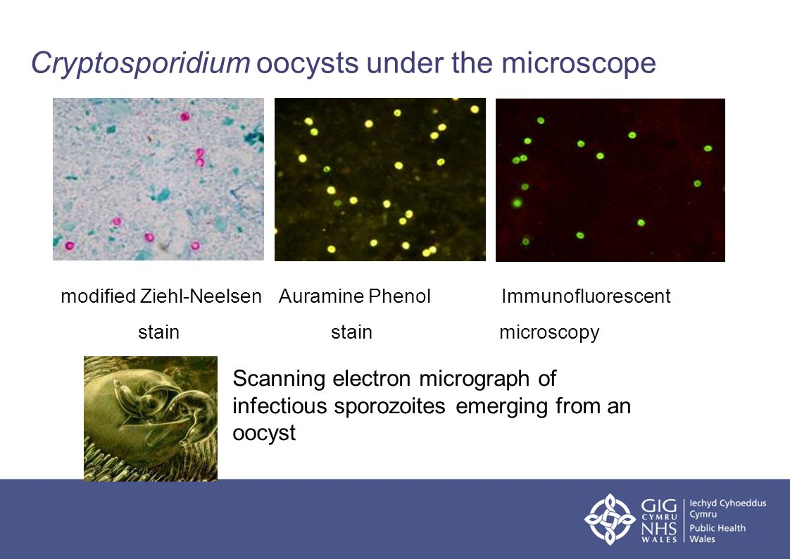
Cryptosporidium Oocyst Under Microscope | When viewed under the microscope, oocysts will appear pink/red in color. Cryptosporidium lives in the intestine of infected humans and animals. Methods for the detection of cryptosporidium oocysts and giardia cysts are very similar and can often be the filter is then sent to the laboratory for further analysis under refrigeration and processed as soon as possible. Dispersion and transport of cryptosporidium oocysts from fecal pats under simulated rainfall events. Daily exposures to cryptosporidium oocysts simulated for individual consumers under conditions consistent with an outbreak (gale and stanfield.
Cryptosporidium oocyst are typically between 3µm and 6µm in size. It spreads through food and water. Cryptosporidium oocysts are environmentally resilient and able to survive for months under suitable conditions 23 and are resistant to chlorination 24. Epa calls for removal or inactivation of 99.9% of cryptosporidium oocysts in drinking water. Letrazuril, an analogue of diclazuril with greater bioavailability, is under study.
All msr microfilters and purifiers meet this standard, including the. Bioaccumulation and elimination of cryptosporidium parvum oocysts in experimentally exposed eastern oysters. Cryptosporidium, sometimes informally called crypto, is a genus of apicomplexan parasitic alveolates that can cause a respiratory and gastrointestinal illness (cryptosporidiosis) that primarily involves watery diarrhea (intestinal cryptosporidiosis) with or without a persistent cough. It is important to specify this request where infection is suspected. Epa calls for removal or inactivation of 99.9% of cryptosporidium oocysts in drinking water. It is a microscopic parasite section of calf intestine infected with crytosporidium parvum and crypto oocysts in fecal smear. Cryptosporidiosis is often diagnosed by finding oocysts in fecal samples from affected animals. Cryptosporidium lives in the intestine of infected humans and animals. Some people may confuse cryptosporidiosis for cryptococcus, as both sometimes go under the name crypto. cryptococcus is a type of invasive fungus that can cause cryptococcosis. Alternative methods for locating objects under microscope have also been demonstrated, including using multiple cameras and calculating location. The main sources of contamination, particularly to surface waters, are from cattle and depending on the quality of the water under analysis, the concentrate volume from the above procedures can vary greatly. Common sense would suggest that a 1µm rated filter would. An endogenous phase of development in microvilli of epithelial surfaces.
Light microscope observations confirmed that relatively often only three sporozoites were released; It is important to specify this request where infection is suspected. Cryptosporidium oocysts have been found on the surface of fresh, raw vegetables obtained from retail markets (ortega et al., 1991 table 16.1. Common sense would suggest that a 1µm rated filter would. Cryptosporidium is a genus of protozoan pathogens which is categorized under the phylum apicomplexa.

Food samples are rarely tested for cryptosporidium oocysts or giardia cysts. Common sense would suggest that a 1µm rated filter would. Sporozoites are sometimes visible inside the oocysts, indicating that sporulation has occurred. Alternative methods for locating objects under microscope have also been demonstrated, including using multiple cameras and calculating location. This is expected, because only a small proportion of the icr concentrates was examined under the microscope, whereas composite samples were used in ims. After the oocysts are purified, they are stained with fluorescent antibodies and detected under the epifluorescence microscope. However, cryptosporidium is extremely resistant to chlorine and chloramines and even in combination the inactivation of cryptosporidium under drinking water. It is a microscopic parasite section of calf intestine infected with crytosporidium parvum and crypto oocysts in fecal smear. Cryptosporidium, sometimes informally called crypto, is a genus of apicomplexan parasitic alveolates that can cause a respiratory and gastrointestinal illness (cryptosporidiosis) that primarily involves watery diarrhea (intestinal cryptosporidiosis) with or without a persistent cough. It spreads through food and water. When viewed under the microscope, oocysts will appear pink/red in color. The dose that can infect humans is low, and a number of waterborne disease outbreaks caused by this protozoan have occurred in the united states, most notably in milwaukee. It is important to specify this request where infection is suspected.
It is important to specify this request where infection is suspected. Daily exposures to cryptosporidium oocysts simulated for individual consumers under conditions consistent with an outbreak (gale and stanfield. Cryptosporidium oocysts are environmentally resilient and able to survive for months under suitable conditions 23 and are resistant to chlorination 24. Two morphofunctional types of oocysts. The fourth one either left the oocyst later arrowood mj, sterling cr (1987) isolation of cryptosporidium oocysts and sporozoites using discontinuous sucrose and isopycnic percoll gradients.
Oocysts are rounded and measure 4.2 to 5.4 µm in diameter. Dispersion and transport of cryptosporidium oocysts from fecal pats under simulated rainfall events. The main sources of contamination, particularly to surface waters, are from cattle and depending on the quality of the water under analysis, the concentrate volume from the above procedures can vary greatly. Light microscope observations confirmed that relatively often only three sporozoites were released; Oocysts (pink arrows) in wet mount. Letrazuril, an analogue of diclazuril with greater bioavailability, is under study. Cryptosporidium is a genus of protozoan pathogens which is categorized under the phylum apicomplexa. Two morphofunctional types of oocysts. Survival of cryptosporidium parvum oocysts under various environmental pressures. All msr microfilters and purifiers meet this standard, including the. Alternative methods for locating objects under microscope have also been demonstrated, including using multiple cameras and calculating location. Entamoeba histolytica cyst under microscope at 40x. Cryptosporidium oocysts were purchased from a commercial source (see the table of materials).
When viewed under the microscope, oocysts will appear pink/red in color cryptosporidium oocyst. Cryptosporidium oocysts are environmentally resilient and able to survive for months under suitable conditions 23 and are resistant to chlorination 24.
Cryptosporidium Oocyst Under Microscope: Dispersion and transport of cryptosporidium oocysts from fecal pats under simulated rainfall events.
0 comments:
Post a Comment